NABH Logo-1

Breast Surgery

DEPARTMENTS

Breast Surgery

Top-Rated Breast Surgeon Hospital in Pimpri Chinchwad

Breast Diseases and Cancer

Anatomy of the Breast

The breast is composed of glandular tissue, fatty tissue, connective tissue, blood vessels, and lymphatic vessels. The primary functional unit of the breast is the lobule, which produces milk, and the ducts that carry milk to the nipple. Surrounding these structures are fatty tissues, which give the breast its shape and size. The breast tissue extends to the armpit, which contains lymph nodes, a key area for the spread of disease.

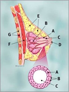
          A –  Lactiferous Duct

          B –  Lactiferous Glands

          C –  Lactiferous Sinus

          D –  Nipple

          E –  Fat Lobules

          F –  Pect. Major

                                                              G –  Ribs and Intercostal Muscles

             A –  Cells lining Ducts

             B –  Basement Membrane

             C –  Duct Lumen

Symptoms of Breast Diseases

  • Lumps or mass in the breast
  • Pain (mastalgia)
  • Nipple discharge (clear, milky, or bloody)
  • Changes in breast size or shape
  • Skin changes (dimpling, redness, swelling)
  • Nipple inversion or changes in the nipple area

[A] Benign (Non-Cancerous) Breast Diseases

  1. Mastalgia (Breast Pain)
    Mastalgia is common and can be cyclical (related to the menstrual cycle) or non-cyclical. Cyclical mastalgia is associated with hormonal changes, while non-cyclical mastalgia could be due to trauma, infections, or musculoskeletal causes.
  2. Nipple Discharge
    Nipple discharge can be due to benign conditions, like duct ectasia, infections, or hormonal fluctuations. However, bloody or spontaneous discharge warrants further investigation to rule out cancer.
  3. Diseases of Lactating Breast
    Lactating women can develop specific issues like milk stasis, blocked ducts, and infections, which can lead to complications like mastitis or breast abscess.
  4. Mastitis
    • Non-Granulomatous Mastitis: Often caused by bacterial infections, this results in inflammation, pain, redness, and swelling. It is commonly seen in breastfeeding women.
    • Granulomatous Mastitis: A rare condition that involves chronic inflammation, possibly due to autoimmune reactions. It may present with firm lumps, abscesses, and fistulas.
  5. Lumps in the Breast


    • Fibroadenoma: A common benign breast tumor that is smooth, mobile, and painless.
    • Cysts: Fluid-filled sacs that can cause discomfort but are generally benign.
    • Cystosarcoma Phyllodes (Phyllodes Tumor): A rare type of breast tumor that can be benign or malignant. It typically grows quickly and may recur after removal.

[B] Breast Cancer

  1. Risk Factors
    • Genetics: Family history of breast cancer, especially if associated with mutations in the BRCA1 and BRCA2 genes.
    • Hormonal Factors: Early menarche, late menopause, use of hormone replacement therapy, and not having children or having the first child after age 30.
    • Lifestyle Factors: Obesity, alcohol consumption, and a sedentary lifestyle are associated with increased risk.
    • Previous Radiation Exposure: Especially in younger individuals for conditions like Hodgkin’s lymphoma.
  2. Signs & Symptoms   


    • A lump or thickening in the breast
    • Changes in the size, shape, or appearance of the breast
    • Nipple retraction or pain
    • Skin dimpling or redness
    • Unexplained nipple discharge (especially bloody)
    • Swollen lymph nodes under the arm
  1. Investigations
    • Mammography: The most common screening tool, especially in women over 40.
    • Ultrasound: Useful for differentiating between solid masses and cysts, especially in younger women.
    • MRI: Often used for high-risk patients or when mammogram/ultrasound results are inconclusive.
    • Biopsy: Tissue sampling is essential for diagnosing cancer and determining its type.
  2. Treatment
    • Surgery: Options include lumpectomy (removal of the tumor) or mastectomy (removal of the entire breast).
    • Radiation Therapy: Often used after surgery to kill remaining cancer cells.
    • Chemotherapy: Involves using drugs to destroy cancer cells, especially in advanced or aggressive cases.
    • Hormonal Therapy: For hormone receptor-positive breast cancer, drugs like tamoxifen or aromatase inhibitors may be used to block hormone receptors.
    • Targeted Therapy: Newer treatments target specific cancer cell markers, like HER2-positive breast cancers.

Self-Breast Examination (SBE)

What is it?
Self-Breast Examination is a technique where a woman examines her own breasts for any changes, lumps, or abnormalities. It should be performed once a month, ideally a few days after menstruation ends when breasts are less likely to be swollen or tender.

Importance

  • Early Detection: SBE allows for early detection of breast lumps, changes in size or shape, or skin and nipple changes, all of which could indicate breast cancer.
  • Empowerment: It promotes breast health awareness, encouraging women to notice normal variations and identify potential problems early, prompting medical consultation.

SBE is not a substitute for professional screening methods like mammography but plays a complementary role in breast health management.

If you have any questions or concerns about breast surgery or would like to schedule a consultation, please do not hesitate to consult our Expert Dr. Deepa Kulkarni (General / Laparoscopic / Hernia Surgeon & Breast Specialist ) for a proper diagnosis and treatment plan. We are here to support you every step of the way.